Tag Archives: orthopaedic surgeon in Delhi

carpal-tunnel-syndrome

Carpal Tunnel Operation

Carpal Tunnel Operation duration

The duration of the Carpal Tunnel Operation is approximately 60 minutes. Generally, the patient can go home the same day of the surgery, although it will depend on the indications of the orthopaedic doctor in Delhi.

What is the Carpal Tunnel Operation?

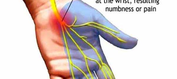
The carpal tunnel syndrome is a condition in which the median nerve is subjected to excessive pressure. The median nerve is located in the wrist and allows the hand to have sensitivity and movement in certain parts. When a person has carpal tunnel syndrome, they often feel numbness, tingling, weakness, or muscle damage to the hand and fingers.

This syndrome can be caused by using tools that vibrate and it is even believed that other activities such as typing, using the mouse, playing an instrument … can cause this inflammation and narrow the carpal tunnel, causing pain.

Other causes can be alcoholism, fractures of the bones, arthritis in the wrist, cyst or tumor in the wrist, infections, obesity, fluids that accumulate during pregnancy and menopause, or rheumatoid arthritis.

It is more common in women than in men and occurs especially in people between 30 and 60 years old.

Surgery Information

What is Carpal Tunnel Release Surgery?

It is a minimally invasive surgical procedure in which the ligament that exerts pressure on the nerve is cut. Local anaesthesia is used and the patient leaves the hospital the same day. At home, the patient is advised to make hand and wrist movements to avoid stiffness and swelling.

The surgical intervention has a very short duration, about 15 minutes.

The use of a special wristband may be indicated during the first three weeks. Of course, overexertion and forced wrist postures should be avoided.

Normally it is a very effective surgery, but it will depend on each patient. Full recovery can take about two months.

After the operation you may feel discomfort or swelling, for this, the orthopaedic in Delhi will prescribe the indicated medication.

Full recovery usually occurs after these 4 or 6 months. This intervention is usually very effective. The innovative techniques used make possible a faster recovery and fewer complications in surgery.

Do not hesitate to go to a consultation with the orthopaedic surgeon in Delhi if you think you may have this problem.

What symptoms does carpal tunnel syndrome have?

Some of the most common symptoms are:

  • The clumsiness of the hand when grasping objects.
  • Numbness and tingling in the thumb and the next two or three fingers. It can also be felt in the palm of the hand.
  • Pain from hand to elbow.
  • Pain in the hand or wrist.
  • Problems with coordination in fine movements.
  • Atrophy of the muscle below the thumb.
  • Difficulty loading bags.
  • Weakness in one or both hands.

What is the initial treatment?

When this syndrome appears, your orthopaedic doctor in Dwarka will advise you to wear a splint at night, avoid sleeping on your wrists and apply cold or heat to the affected area. Additionally, nonsteroidal anti-inflammatory drugs (NSAIDs) or corticosteroid injections may be prescribed to relieve symptoms.

When none of these treatments works, carpal tunnel release surgery can be used. In the long term, surgical treatment is the only solution for carpal tunnel syndrome.

Femur-Fracture

Open Reduction and Internal Fixation of Femur Fracture

What is an open reduction and internal fixation of the femur fracture?

Open reduction and internal fixation is a surgery used to heal and stabilize a broken bone. You may need this procedure to treat your broken femur (thigh bone).

The femur is the large bone in the upper leg. Various types of trauma can damage this bone, causing it to fracture into two or more pieces. This can occur in the part of the femur near the knee, near the middle of the femur, or in the part of the femur that is part of the hip joint. In some types of femur fractures, your femur has ruptured, but its parts still line up correctly. In other types of fractures (displaced fractures), trauma moves the bone fragments out of alignment.

Usually, you need open reduction and internal fixation to get your bones back into place and help them heal when the femur fractures. During an open reduction, the orthopaedic surgeons replace their bone pieces during surgery, so that they are back in their correct alignment. This is in contrast to a closed reduction, in which an orthopaedic doctor in Delhi physically moves the bones into place without surgically exposing them.

Internal fixation can be defined as a method of physically reconnecting your bones. This could include special orthopaedic screws, plates, rods, wires, or nails that your orthopaedic surgeon in Delhi places inside your bones to put them in the correct position. This prevents your bones from healing abnormally. For a fracture of the long and middle part of your femur, your surgeon may insert a long metal rod through the middle of your bone. The complete operation is normally performed while you are asleep under general anaesthesia.

Why might you need open reduction and internal fixation of the femur fracture?

Some medical conditions can increase the chance of breaking your femur. For example, if you are an older adult, osteoporosis increases the risk of fracturing your femur. Your femur may also be expected to rupture if you have bone cancer. Auto accidents, firearm injuries, sports-related injuries, and falls are common sources of trauma that can cause a femur fracture. A direct blow to the hip can break the part of the femur associated with the hip joint.

Most people with a fractured femur need some type of surgery, usually open reduction and internal fixation. Your broken femur may not heal properly without surgery. Open reduction and internal fixation can put your bones back in their proper settings. This significantly increases the chance that your bone will heal properly. Your orthopaedic doctor in Delhi may recommend nonsurgical treatment for a very young child or for people with other medical conditions that make surgery more dangerous.

You may need open reduction and internal fixation for a fracture that occurs anywhere in your femur, including the part that is part of your hip joint. In a “broken hip,” it is usually a part of your femur that breaks, and not part of the hip bone itself.

elbow-fracture

What to do with an Elbow Fracture

They are frequent in children and can become complex.

The fractures of the elbow include a wide spectrum of injuries whose treatment ranges from a brief period of immobilization and immediate rehabilitation to complex surgery and long periods of rehabilitation.

Dr. Ashu Consul an elbow and shoulder specialist traumatologist, orthopaedic surgeon in Delhi, indicates that when faced with a traumatic elbow injury, the first thing to do is apply ice, immobilize with a sling and consult an emergency or trauma service to Clinically and imaging the lesion.

“In those minor fractures, for example, fractures of the radius dome without displacement, a short immobilization period of 7-10 days is sufficient, and then early mobilization begins, sometimes assisted with kinesiology depending on the injury, ”orthopaedic in Delhi explains.

Above all, in children, elbow fractures are frequent, especially those that involve the distal humerus says the orthopaedic doctor in Delhi. “Its management ranges from plaster for 3 weeks in those nondisplaced fractures, and surgery with reduction (alignment of the fracture) and needle fixation in those displaced fractures,” he indicates.

There are severe elbow injuries that occur in higher energy trauma and these involve severe bone or soft tissue (ligament) injury. “These types of injuries may require a complex plate and screw fixation surgeries and long periods of rehabilitation,” says Dr. Consul.

The elbow is a joint in which stiffness is frequently presented as a complication, which is why its management must be carried out by specialists, both trauma and kinesiologists, he emphasizes.

Full and definitive recovery from a complex elbow fracture can take 6 to 8 months.

Disc-Herniation

Everything you need to know about Disc Herniation

Disc herniation means the movement of discs in the spine. As a result of this condition, intense back pain occurs in that area.

Depending on the area of ​​the back where they occur, the most common types of herniated disc are lumbar and cervical.

Lumbar disc herniation occurs when the movement of the intervertebral disc has occurred at the lumbar level. The most affected are the last two lumbar discs, L4 and L5, due to the fact that they support most of the body weight. The most affected nerves are the sciatic and femoral.

Cervical disc herniation occurs when the displacement of the intervertebral disc has occurred in the upper part of the spine.

Causes

Disc herniation is most often the result of gradual wear and tear related to aging, called pulpal nucleus degeneration. As a person gets older, the discs lose some of their fluid content. This makes them less flexible and more prone to cracking or breaking, even in a small movement or twist.

Predisposition to sedentary lifestyle, prolonged sitting, increased exertion after a longer period of rest, lifting weights incorrectly, but also spinal cord injuries are among the causes of disc herniations, explains the orthopaedic in Delhi.

Risk factors

Weight. Excessive weight causes extra stress on the lower back discs.

Occupation. People with high-intensity jobs are at higher risk of developing spinal problems. Lifting, pulling, or pushing weights, repeated movements, or lateral bending and twisting may increase the risk of a herniated disc.

Genetics. Some people inherit a predisposition to the development of a herniated disc.

Symptoms

The symptoms of a herniated disc differ depending on the area in which it occurs.

In the case of lumbar disc herniation, there is pain in the thighs and buttocks, which can radiate to the leg below the knee. Pain may be accompanied by tingling, numbness and tenderness in the limbs and affected areas.

In the case of cervical hernia, the pain manifests itself mainly in the shoulders and arms. There is pain in the neck region, with the impossibility of turning the head or making sudden movements.

The pain may be accompanied by tingling, numbness and a general state of weakness. In both cases, the pain is continuous and not throbbing.

Treatment

Disc herniation treatment should not be delayed because the pain subsided at some point. Lack of treatment can worsen the situation, causing chronic pain or permanent injuries that affect mobility, says the orthopaedic surgeon in Delhi.

Physiotherapy is the most common methods of treating a herniated disc. In severe cases, when the pain persists even after 5-6 weeks of alternative treatment, surgery is resorted to.

The operation consists of removing the fragments of the disc or even the entire herniated disc that compresses the spinal nerves.

Patella-Dislocation

Dislocations and Fractures of the Patella

Disorders of the patella are quite common conditions, they can occur on a normal knee or previously affected by congenital diseases. From a clinical point of view, there are spontaneous or aggravated pain due to flexion of the leg, they may have variable intensity.

When the knee is inspected, the displaced patella is observed and in its place, a depression occurs. Hydrarthrosis can accompany the condition, without being important.

The treatment of luxation is orthopedic or compressive bandage can be used. In severe cases, it may be possible to reach the patellectomy. Recovery through physical therapy is mandatory.

The fractures of the patella can occur both directly and indirectly, by an exaggerated flexion of the calf.

Clinically, pain occurs, joint swelling, hemarthrosis, functional impotence. At the touch of the knee, the displaced fragments are noticed.

The treatment of fractures may vary depending on the severity. It goes from rest to bed, compressive bandage, to gypsum cast, patellectomy. The walk can be resumed after about three weeks.

The recovery of the fractured knee is made from the period of immobilization: treating inflammation, ensuring lymphatic drainage, maintaining muscle tone, recovering after the end of immobilization by passive exercises done by kinetotherapist in water, but also by active movements, reinventing the gait at the beginning with carriage and then with a cane., reconfiguration to the orthostatic position, toning of the muscles, joint mobilization and regaining the amplitude of flexion, are the steps that will have to be followed.

Prophylactic treatment is a very important one, in that the sudden movements at the knee level will be avoided by the performance athletes but also by the elderly and children who have increased sensitivity at this level.