Category Archives: Blogs

arthroscopy in Delhi

Knee Ligament Operation or Arthroscopy

Cross Ligament Operation

Arthroscopy is a minimally invasive technique, which carries a 50% less chance of infection and a faster recovery compared to the classical technique.

The reconstruction of the cruciate ligaments also called knee arthroscopy in Delhi. It is a surgery to rebuild the ligament in the centre of the knee. This ligament, the anterior cruciate ligament, holds the tibia in place. Its breakage can cause that during physical activity, the knee does not move correctly.

Injuries to the cruciate ligament cause instability in the knee. And in the long run, this increases the likelihood of a meniscus tear. The cruciate ligament reconstruction is also recommended when knee pain, when it becomes impossible to sport or other daily activities, when other ligaments are damaged or when there is a meniscal tear.

Information about surgery

What does Cruciate Ligament Surgery consist of?

The knee arthroscopy operation consists of replacing the anterior cruciate ligament with tissue from the patient’s own body and is usually taken from a patella tendon or a hamstring. During the procedure, orthopaedic surgeon in Delhi inserts a camera into the knee through a small incision. Through other small incisions around the knee, the rest of the medical instruments are introduced. The old ligament is removed and the new ligament is fixed with screws or other devices to hold it in place. The advantage of this technique over others is the speed with which the patient can return to a normal life. The type of anaesthesia used for this intervention is general or epidural.

How is the postoperative period?

Typically, the patient leaves the hospital the day after surgery (depending on each patient) and must use crutches between the first and fourth week. It is advisable to move your knee immediately after surgery to prevent stiffness.

Once at home, the patient will need the help of a friend or relative to carry out daily activities. It is not recommended to take the weight, or to bend the knee too much until the ligament is well seated.

After the operation, it is usually necessary for the patient to follow a rehabilitation program for 4 to 6 months. This routine can help you regain normal movement and strength in your knee.

carpal-tunnel-syndrome

Carpal Tunnel Operation

Carpal Tunnel Operation duration

The duration of the Carpal Tunnel Operation is approximately 60 minutes. Generally, the patient can go home the same day of the surgery, although it will depend on the indications of the orthopaedic doctor in Delhi.

What is the Carpal Tunnel Operation?

The carpal tunnel syndrome is a condition in which the median nerve is subjected to excessive pressure. The median nerve is located in the wrist and allows the hand to have sensitivity and movement in certain parts. When a person has carpal tunnel syndrome, they often feel numbness, tingling, weakness, or muscle damage to the hand and fingers.

This syndrome can be caused by using tools that vibrate and it is even believed that other activities such as typing, using the mouse, playing an instrument … can cause this inflammation and narrow the carpal tunnel, causing pain.

Other causes can be alcoholism, fractures of the bones, arthritis in the wrist, cyst or tumor in the wrist, infections, obesity, fluids that accumulate during pregnancy and menopause, or rheumatoid arthritis.

It is more common in women than in men and occurs especially in people between 30 and 60 years old.

Surgery Information

What is Carpal Tunnel Release Surgery?

It is a minimally invasive surgical procedure in which the ligament that exerts pressure on the nerve is cut. Local anaesthesia is used and the patient leaves the hospital the same day. At home, the patient is advised to make hand and wrist movements to avoid stiffness and swelling.

The surgical intervention has a very short duration, about 15 minutes.

The use of a special wristband may be indicated during the first three weeks. Of course, overexertion and forced wrist postures should be avoided.

Normally it is a very effective surgery, but it will depend on each patient. Full recovery can take about two months.

After the operation you may feel discomfort or swelling, for this, the orthopaedic in Delhi will prescribe the indicated medication.

Full recovery usually occurs after these 4 or 6 months. This intervention is usually very effective. The innovative techniques used make possible a faster recovery and fewer complications in surgery.

Do not hesitate to go to a consultation with the orthopaedic surgeon in Delhi if you think you may have this problem.

What symptoms does carpal tunnel syndrome have?

Some of the most common symptoms are:

  • The clumsiness of the hand when grasping objects.
  • Numbness and tingling in the thumb and the next two or three fingers. It can also be felt in the palm of the hand.
  • Pain from hand to elbow.
  • Pain in the hand or wrist.
  • Problems with coordination in fine movements.
  • Atrophy of the muscle below the thumb.
  • Difficulty loading bags.
  • Weakness in one or both hands.

What is the initial treatment?

When this syndrome appears, your orthopaedic doctor in Dwarka will advise you to wear a splint at night, avoid sleeping on your wrists and apply cold or heat to the affected area. Additionally, nonsteroidal anti-inflammatory drugs (NSAIDs) or corticosteroid injections may be prescribed to relieve symptoms.

When none of these treatments works, carpal tunnel release surgery can be used. In the long term, surgical treatment is the only solution for carpal tunnel syndrome.

Femur-Fracture

Open Reduction and Internal Fixation of Femur Fracture

What is an open reduction and internal fixation of the femur fracture?

Open reduction and internal fixation is a surgery used to heal and stabilize a broken bone. You may need this procedure to treat your broken femur (thigh bone).

The femur is the large bone in the upper leg. Various types of trauma can damage this bone, causing it to fracture into two or more pieces. This can occur in the part of the femur near the knee, near the middle of the femur, or in the part of the femur that is part of the hip joint. In some types of femur fractures, your femur has ruptured, but its parts still line up correctly. In other types of fractures (displaced fractures), trauma moves the bone fragments out of alignment.

Usually, you need open reduction and internal fixation to get your bones back into place and help them heal when the femur fractures. During an open reduction, the orthopaedic surgeons replace their bone pieces during surgery, so that they are back in their correct alignment. This is in contrast to a closed reduction, in which an orthopaedic doctor in Delhi physically moves the bones into place without surgically exposing them.

Internal fixation can be defined as a method of physically reconnecting your bones. This could include special orthopaedic screws, plates, rods, wires, or nails that your orthopaedic surgeon in Delhi places inside your bones to put them in the correct position. This prevents your bones from healing abnormally. For a fracture of the long and middle part of your femur, your surgeon may insert a long metal rod through the middle of your bone. The complete operation is normally performed while you are asleep under general anaesthesia.

Why might you need open reduction and internal fixation of the femur fracture?

Some medical conditions can increase the chance of breaking your femur. For example, if you are an older adult, osteoporosis increases the risk of fracturing your femur. Your femur may also be expected to rupture if you have bone cancer. Auto accidents, firearm injuries, sports-related injuries, and falls are common sources of trauma that can cause a femur fracture. A direct blow to the hip can break the part of the femur associated with the hip joint.

Most people with a fractured femur need some type of surgery, usually open reduction and internal fixation. Your broken femur may not heal properly without surgery. Open reduction and internal fixation can put your bones back in their proper settings. This significantly increases the chance that your bone will heal properly. Your orthopaedic doctor in Delhi may recommend nonsurgical treatment for a very young child or for people with other medical conditions that make surgery more dangerous.

You may need open reduction and internal fixation for a fracture that occurs anywhere in your femur, including the part that is part of your hip joint. In a “broken hip,” it is usually a part of your femur that breaks, and not part of the hip bone itself.

elbow-fracture

What to do with an Elbow Fracture

They are frequent in children and can become complex.

The fractures of the elbow include a wide spectrum of injuries whose treatment ranges from a brief period of immobilization and immediate rehabilitation to complex surgery and long periods of rehabilitation.

Dr. Ashu Consul an elbow and shoulder specialist traumatologist, orthopaedic surgeon in Delhi, indicates that when faced with a traumatic elbow injury, the first thing to do is apply ice, immobilize with a sling and consult an emergency or trauma service to Clinically and imaging the lesion.

“In those minor fractures, for example, fractures of the radius dome without displacement, a short immobilization period of 7-10 days is sufficient, and then early mobilization begins, sometimes assisted with kinesiology depending on the injury, ”orthopaedic in Delhi explains.

Above all, in children, elbow fractures are frequent, especially those that involve the distal humerus says the orthopaedic doctor in Delhi. “Its management ranges from plaster for 3 weeks in those nondisplaced fractures, and surgery with reduction (alignment of the fracture) and needle fixation in those displaced fractures,” he indicates.

There are severe elbow injuries that occur in higher energy trauma and these involve severe bone or soft tissue (ligament) injury. “These types of injuries may require a complex plate and screw fixation surgeries and long periods of rehabilitation,” says Dr. Consul.

The elbow is a joint in which stiffness is frequently presented as a complication, which is why its management must be carried out by specialists, both trauma and kinesiologists, he emphasizes.

Full and definitive recovery from a complex elbow fracture can take 6 to 8 months.

hip-replacement in Delhi

FAQ about hip replacement

Orthopaedic doctor in Delhi has always recommended avoiding joint wear and tear to maintain movement, hopefully throughout life. This need has been so pressing for humanity that more than 100 years ago, surgeries were already performed in which tissues or elements such as metal sheets were interposed to hold the bones in place.

However, the first complete joint change was not made until 1938, when the first hip was replaced in the United States; a procedure that was improving until in 1960 the Englishman John Charnley laid the foundations for doing transplants of this joint in a systematic way.

What is it and what is changed?

Total hip replacement is the exchange of all the components of the joint for an artificial implant called a prosthesis. The hip has two main parts: a cavity in the pelvis called the acetabulum and the head of the femur, and they are the ones that are replaced.

Why do you have to replace a hip?

Because the cartilage wears out and the bones begin to touch each other, which produces pain and deterioration, configuring a condition called osteoarthritis. This is reached by problems in development, some diseases, overweight and the wear and tear of age. The hip also needs to be replaced in cases of traumatic fractures or destruction.

And works?

Of course. By replacing it, the person can lead a normal life and when measuring its results in terms of quality of life, it is one of the most favorable procedures, which earned it the most important surgery of the last century.

How long is the procedure?

About an hour. It is a safe, reproducible and long-lasting procedure.

How is the recovery?

This starts from the first day of surgery with movements in the feet and knees. The next day, the person can already support in the middle of a rehabilitation program and at the following 6 or 8 weeks the person can resume normal activity.

How does a person feel with a different material in the body?

Pain relief from the first day is the first feeling that people refer to and eventually they say they feel something strange, but not annoying in those places.

Do the children have surgery?

No. It is a procedure that requires skeletal maturity. So it can be operated at the end of adolescence.

What complications does it have?

Although it is a very safe procedure, it is not without infections or dislocations that occur in a very low percentage.

What is the future of prosthetics?

Nowadays, it is sought that they be less invasive, that they provide greater mobility and that they be made with more resistant and durable materials, so that the objective of the surgery is to recover all activities, including recreational and sports activities.

hip-joint-pain

The Main Causes That Influence Hip Pain

When you tell your orthopaedic in Delhi that your hip is hurting, the first thing the doctor needs to do is confirm that the balance is the problem.

Women can say they have hip pain, but they can also say that they have pain in the upper thigh or upper buttock.

These hip pains may be faced with minor back pain. Hip pain can be felt very often in the area of ​​the abdomen or the outer part of the hip over which its joint is located.

Causes of hip pain

If a female patient complains of hip pain, the orthopaedic in Dwarka firstly considers the patient’s age and level of activity. Among the most common causes of hip pain in women, we mention:

  1. Arthritis. The most common causes of chronic hip pain in women are arthritis, especially osteoarthritis, which affects many people as they get older. Arthritis pain is most often felt in the front of the thigh or in the abdomen area, due to stiffness or swelling.
  2. Hip fractures. Hip fractures are common in older women, especially those with osteoporosis (decreased bone density). Symptoms of a hip fracture include severe pain.
  3. Tendonitis and bursitis. Many tendons around the hip connect the muscles with each other. These tendons can easily become inflamed if you overload them or participate in intense physical activity. One of the most common causes of tendonitis in the hip joint, especially for athletes, is the iliotibial tract syndrome. Another common cause of hip pain in women is exchange. There are some liquid bags in the bony side of the hip, called bursa pillows. Like tendons, these bags may become inflamed from irritation or over-stress, causing pain every time the hip joint moves.
  4. Hernia. In the abdomen area, the groin and femoral hernias – sometimes referred to as sports hernias – can cause anterior (frontal) pain in the woman’s hip. Pregnant women may be sensitive to groin hernias due to the pressure added to the wall of their abdomen.
  5. Gynecological problems. Hip pain in women can also have gynecological causes. It is important not to just realize that the pain can be caused by arthritis, bursitis or tendonitis. Depending on the age or other health problems, hip pain may also come from another system. Endometriosis can cause pelvic sensitivity, so some women describe this problem as hip pain.

Back pain or spinal pain can also be mentioned and felt around the buttocks or hip. Sciatica can cause pain in the back of the hip – sciatica pain can start in the lower back and reach the buttocks and even the legs.

Arthritis is a common cause of hip pain and mobility change. More than a quarter of older adults develop coxarthrosis pain, which threatens mobility – slower walking, difficult stairs. The term “arthritis” is covered under a number of different conditions, including osteoarthritis and inflammatory diseases, such as rheumatoid arthritis and psoriatic arthritis. If you find out what kind of arthritis you have, you must be able to successfully reduce hip pain. And finding out the type of arthritis is a solution that reduces your hip pain. The most common type of arthritis is osteoarthritis. Osteoarthritis can also result from a common injury, sometimes called traumatic arthritis.

Factors that increase the risk of osteoarthritis include:
1. Aging;
2. Obesity;
3. Deterioration or various injuries in the joint;
4. Structural problems with the joint;
5. Rheumatoid arthritis.

Symptoms of osteoarthritis develop slowly, starting with stiffness or pain in one or both bones of the hip and eventually becoming painful enough to prevent various normal activities (walking or climbing stairs).

Rheumatoid arthritis can also be a problem that causes pain. Inflammation may occur due to an abnormality of the immune system. Rheumatoid arthritis is a chronic disease that can affect the entire body, not just the hips. It starts with a swelling of the mucosa of the joints, called the synovial mucosa and progresses to the deterioration of bones and cartilage. The cause of rheumatoid arthritis is not fully understood, although an abnormal response of the immune system to the body contributes to the disease.

Symptoms of rheumatoid arthritis also include:
a. Pains that increase slightly in intensity in the symmetrical joints;
b. Swelling of the affected joints;
c. Fatigue;
d. morning rigidity;
e. Pain after a period of stay;
f. Weakness;
g. Muscle pain ;
h. Anemia.

Early diagnosis of rheumatoid arthritis is critical to maintaining the quality of life. Medications can slow the progression of this disease. A less common type of arthritis that causes joint pain is psoriatic arthritis, a complication of psoriasis, inflammatory skin disease.

Strategies to ease discomfort:

  • Lose weight if you are overweight in order to ease the weight of the joints;
  • Try analgesics, but be careful about recommended doses or warnings;
  • Ask your doctor for more information about common supplements such as glucosamine and chondroitin.
  • Work with a physical therapist to do stretching and flexibility exercises;
  • Use ice and heat to relieve painful areas.

If your mobility is impaired, ask your orthopaedic in West Delhi if you would like the assistance of a support device to help you with walking or other daily activities. Discuss in detail the options for prescription drugs, injections, or surgeries with the orthopaedic surgeon in Delhi so that a treatment plan is tailored to your needs.

Managing hip pain

Things you can do to better manage your hip pain:

  1. Exercises – do exercises in the morning to put your muscles to work, to be active. Press on your ankles and lift your buttocks off the floor as you tighten your abdominal muscles. Keep your knees aligned with your ankles and follow a straight line from your knees to your shoulders, without bending your back. Stay in that position for three to five seconds. Start with a set of ten exercises.
  2. Use ice – against arthritis or bursitis ice can be used, thus reducing inflammation and helping against hip pain. Use an ice pack, wrap it in a towel and place it where you feel the pain.
  3. Use heat for arthritis – warming an arthritic balance with a hot shower or bath can relieve pain. Do not use heat, if hip pain is caused by the stock market.
  4. Strengthening the thighs – this is another muscle group that contributes to supporting the hips. Lie on your back, place a ball between your knees and tighten. And a hard pillow or Pilates ring will help. Start with a set of ten repetitions and proceed with approximately up to three sets. Then strengthen your thighs on the outside.
  5. Work out in the water – swimming or aerobics in water are wonderful exercises for the hip joints. Exercises in water allow you to strengthen your muscles without putting pressure on your joints.
  6. Avoid activities that have a high impact – jumps can cause hip pain to worsen. Walking is a better choice.
  7. Weight loss – if you have osteoarthritis in the hip that results from cartilage wear, losing even a few pounds can help against joint and hip pain.
  8. Listen to your body – if you have arthritis or tummy tuck, you may have noticed that exercise can help relieve pain. If the balance starts to hurt during certain exercises and persists longer, this is a sign that you need rest and that you should rest. It is normal to feel pain one day after exercise, but the pain should not persist or become worse. Also, if you experience sharp pain, stop your activity and talk to a doctor or a physiotherapist in Delhi.

If you undergo major hip replacement surgery in Delhi, it is very important to prepare yourself in advance to increase your chances of an optimistic outcome and reduce hip pain. The most important thing you can do before surgery is to make sure you are on the same wavelength as the orthopedic surgeon. You need a doctor to talk to and gain confidence in, so you get a sense of well-being. Before replacing your balance, you will be evaluated by your orthopaedic surgeon in Dwarka to make sure you are healthy enough for the operation.

This evaluation could include an electrocardiogram, chest x-ray, and various blood tests. Your doctor will use this information to determine if you have an increased risk of complications. Note that certain herbal medicines, including supplements, may increase the risk of bleeding and other serious complications during surgery. So make sure that both your orthopaedic surgeon in West Delhi and you are aware of all the medicines, herbal supplements you are administering.

Balance replacement is a major operation and there is the possibility of needing a blood transfusion during or after the operation. However, you can start donating blood about four weeks before surgery. Inform yourself by asking your orthopedic in Delhi what you should do about how much blood can be stored safely before surgery.

To produce more red blood cells (this requires an injection once a week for about a month before surgery), you can also take other medicines (epoetin alfa). In this way, patients have an extra reserve in terms of blood supply when they undergo surgery.

Men with prostate problems should be checked by a urologist prior to surgery. After you schedule your hip replacement in Delhi, call your dentist. If you have problems with your teeth, you should go for a checkup. Infections from an abscess or other dental infection can spread to the hip, causing serious complications.

Cuboid

Leg Injuries – Cuboid Syndrome

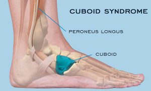
Cuboid syndrome is the result of injuries to the joints and ligaments surrounding the cuboid bone. This is one of the seven tarsal bones of the foot (talus or astragalus, calcaneus, navicular, cuboid and 3 cuneiform bones).

From a medical point of view, the condition is called a cuboid subluxation and occurs when the cuboid bone moves down and out, without being aligned with the calcaneus bone.

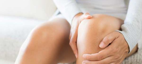
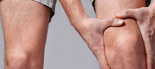
The cuboid syndrome causes pain in the external side of the foot, which can be confused with other conditions.

Clinical picture

Cuboid syndrome encompasses a variety of common symptoms and other conditions, sometimes making it difficult to diagnose. The most common symptoms include:

  • Pain on the outside of the foot, which affects including fingers 4 and 5;
  • Pain that aggravates the standing state, due to body weight ;
  • Painful pain, dry or sharp and acute;
  • Weakness and difficulty in walking and jumping;
  • Redness, inflammation, and sensitivity in the injured area;
  • Walking painkillers, to reduce pain;
  • Reducing the range of movements of the foot and ankle.

Studies have reported that, in general, the cuboid syndrome is not uncommon in the population, but occurs most commonly in athletes and dancers. When the cuboid syndrome is correctly identified and treated, most people experience a complete recovery.

Causes

Cuboid syndrome can have different causes, but the most common are:

  • Overload – this condition, along with injury, is most commonly seen in athletes and dancers. These groups of athletes tend to have intense physical activity, regardless of the presence of pain. Overload injuries tend to develop after long periods of intense activity, such as running;
  • Ankle dislocation – ankle dislocation can also lead to injury to the Tarsus bones of the foot. A specialist study reported that about 40% of people with ankle dislocations or sprains can develop cuboid syndrome;
  • Foot walking in pronation – people who develop this type of walking have a higher risk of having the cuboid syndrome because the muscles of the leg can pull the tarsal bones into abnormal positions;
  • Other causes – climbing, uneven surfaces and sports involving fast and lateral movements, such as tennis, can cause the cuboid syndrome. Wearing shoes of poor quality or inadequate sizes may be another reason.

Also, if you do not take into account the indications of orthopaedic in Delhi regarding rest and recovery from a leg injury, various complications, including cuboid syndrome, may occur.

Diagnostics


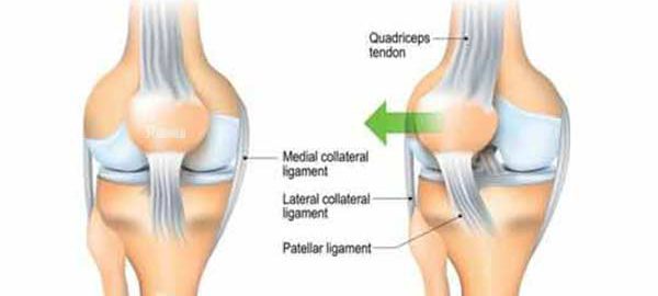
The foot represents a complex, flexible and resistant segment of the body. It contains about 100 muscles, ligaments and tendons, 28 bones and 30 joints.

The complex structure of the foot and the non-specific nature of the pain within the cuboid syndrome make this injury difficult to diagnose.

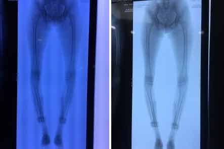
Sometimes, medical imaging techniques such as X-ray or magnetic resonance imaging (MRI) do not identify signs of this condition, even if it is present. The cuboid syndrome may also mimic the symptoms of other foot problems, such as fractures or sprained heels.

In order to establish a correct diagnosis and to find the most effective treatment, the physician must perform a thorough physical examination and examine a person’s medical history.

Treatment options

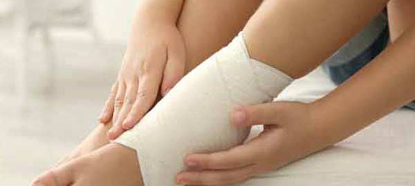
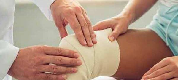
Treating cuboid syndrome always involves rest and elimination of activities that involve putting pressure on the injured leg. Applying ice and an elastic phase, but also raising the foot above the level of the heart on a pillow are other useful methods.

If the pain persists or worsens, the orthopaedic in Dwarka should be consulted urgently. Certain maneuvers performed by the therapists in the field can help to improve or eliminate the symptoms.

These maneuvers are meant to force the bone to return to its original position. However, it should be noted that such maneuvers are not recommended if there are other associated conditions, such as arthritis, bone disease, fractures, circulatory or nerve disorders.

Additional treatments may include the use of orthotics to support and properly align the foot, but also to administer anti-inflammatory drugs to reduce pain and inflammation.

In some cases, leg massage can help to reposition the bone. Surgery is used in rare cases when other treatment options have not worked.

Recovery

The length of time required for recovery from cuboid syndrome depends on several factors, such as:

  • Duration of the treatment until the start of treatment;
  • Cause of cuboid syndrome ;
  • If the initial injury was mild or severe;
  • If the instructions of orthopaedic in West Delhi were taken into account;
  • If the condition has been correctly diagnosed and treated.

Physiotherapy in Dwarka can play an important role in the complete recovery of a person with the cuboid syndrome. Also, this therapy can prevent further complications.

Physical therapy includes:

  • Reconsolidation of the strength and function of the foot;
  • Maintenance of the muscles;
  • Exercises to improve balance.

In some cases, to avoid pressure on the injured leg, the orthopaedic surgeon in Delhi may indicate the use of squats or walking sticks.

Risk factors

The common risk factors for the cuboid syndrome are:

  • Overweight or obesity;
  • Wearing shoes of inappropriate sizes;
  • Wearing pairs of shoes of inferior quality;
  • Practicing physical exercises without adequate heating;
  • Practicing physical activities without taking into account pain and without having sufficient rest periods;
  • Performing activities, such as running or walking on uneven or rough surfaces;
  • Fractures of neighboring bones of the cuboid;
  • Practicing sports such as athletics or ballet;
  • Practicing physical activities that involve repetitive movements and increased impact on the foot;
  • Association of other conditions such as arthritis or osteoporosis.

Perspective

Cuboid syndrome is not a serious condition and can be easily treated by the orthopaedic surgeon in Dwarka, sometimes involving physical therapy. The perspective is usually very good for people with this condition. Following treatment, most people can return to normal activities, with little risk of recurrence.

The orthopedic in Delhi should be notified if persistent pain occurs in the foot before doing any maneuver that could aggravate the situation.

Often there may be other health conditions that mimic cuboid syndrome. Rapid identification and early treatment of health status offer the best chances of recovery.

meniscus lesions of knee joint

Meniscus Lesions of The Knee Joint

The two meniscus can be easily injured by a rotation of the knee during a sustained effort. Partial or total meniscus rupture occurs at an abrupt torsion or thigh rotation, while the foot stays in place (for example, at a sudden turn to hit the tennis ball).

If the rupture is minimal, the meniscus remains connected in the anterior and posterior part of the knee; if the rupture is large, the meniscus may hang from a cartilage filament. The severity of a rupture depends on the location and extent.

Symptoms

In general, pain in the meniscus rupture occurs, especially when the leg is straightened (at the extension of the leg). The pain may be moderate and the patient may continue the activity.

Severe pain occurs when a fragment of the meniscus remains between the femur and tibia. Swelling may occur immediately after injury if the blood vessels are damaged, or a few hours after injury if the joint space is filled with fluid produced in response to inflammation. If the synovial envelope is damaged, it becomes inflamed and produces fluid to protect itself, installing the synovial envelope syndrome.

These changes cause swelling of the knee. Sometimes an untreated injury can become painful after several months or even years later, especially if the knee has been injured a second time. After an injury, the knee may become blocked or weakened. Symptoms of a meniscus lesion may resolve spontaneously, but frequently the symptoms persist and require treatment.

Diagnostic

In addition to the patient’s anamnesis, which describes the onset of pain and swelling, the orthopaedic in Dwarka may perform an x-ray of the affected knee. The examination may include a test in which the doctor flexes the leg, then rotates internally and externally while extending the leg. The pain from this maneuver suggests the meniscus rupture. An imaging magnetic resonance test may be recommended to confirm the diagnosis. Occasionally, the orthopaedic surgeon in Dwarka may use arthroscopy for diagnosis and treatment.

Treatment

If the injury is minimal and the pain and other symptoms disappear, the orthopaedic in Delhi will recommend a program of exercises to strengthen the muscles. Exercises for meniscus disorders are initially performed under the supervision of an orthopaedic in West Delhi or therapist.

The physiotherapist in Dwarka will ensure that the patient performs the exercises correctly and without the risk of a new injury. The following exercises are intended to strengthen the thigh muscles and increase the elasticity:

  • warming the joint by walking on a medical bicycle, then stretching and lifting the leg
  • extension of the foot from the sitting position (a small weight can be placed on the ankle for this exercise)
  • in ventral decubitus, there is performed leg lifting movements
  • exercises in the swimming pool, including the rapid walking with water at the chest level, raising each leg to 90 with the back glued to a wall of the pool.

If the rupture is enlarged, the doctor may perform arthroscopy in Delhi or surgery to assess the severity and repair the lesion. The orthopaedic surgeon in Delhi can suture the meniscus in relatively young patients if the lesion is in a well-vascularized area and the ligaments are intact. Most young athletes will be able to perform vigorous sports activities after meniscus repair.

In elderly patients or if the rupture is in a poorly vascularized area, the doctor will resect a small portion of the meniscus to smooth the surface. In some cases, the orthopaedic surgeon in West Delhi will resect the entire meniscus.

However, degenerative rheumatism, such as osteoarthritis, presents a higher risk of development if the meniscus is completely removed. Researchers are investigating a procedure by which the meniscus is replaced with a meniscus from a corpse (allograft). A meniscus graft is fragile and can shrink and break easily. The researchers tried to replace the meniscus with an artificial one, but the procedure was even less successful than the allograft.

The rehabilitation after the surgical repair of the meniscus lasts for several weeks, and the postoperative activity is more restricted than in the case of the total resection of the meniscus.

However, exercises speed up healing. Regardless of the type of surgical operation, rehabilitation includes gait, flexion and extension exercises of the knee, and exercises for strengthening the thigh muscles.

The best results of treatment for a meniscus lesion are obtained in patients without modifications of the articular cartilage and without lesions of the anterior cruciate ligament intact.

Patella-Dislocation

Dislocations and Fractures of the Patella

Disorders of the patella are quite common conditions, they can occur on a normal knee or previously affected by congenital diseases. From a clinical point of view, there are spontaneous or aggravated pain due to flexion of the leg, they may have variable intensity.

When the knee is inspected, the displaced patella is observed and in its place, a depression occurs. Hydrarthrosis can accompany the condition, without being important.

The treatment of luxation is orthopedic or compressive bandage can be used. In severe cases, it may be possible to reach the patellectomy. Recovery through physical therapy is mandatory.

The fractures of the patella can occur both directly and indirectly, by an exaggerated flexion of the calf.

Clinically, pain occurs, joint swelling, hemarthrosis, functional impotence. At the touch of the knee, the displaced fragments are noticed.

The treatment of fractures may vary depending on the severity. It goes from rest to bed, compressive bandage, to gypsum cast, patellectomy. The walk can be resumed after about three weeks.

The recovery of the fractured knee is made from the period of immobilization: treating inflammation, ensuring lymphatic drainage, maintaining muscle tone, recovering after the end of immobilization by passive exercises done by kinetotherapist in water, but also by active movements, reinventing the gait at the beginning with carriage and then with a cane., reconfiguration to the orthostatic position, toning of the muscles, joint mobilization and regaining the amplitude of flexion, are the steps that will have to be followed.

Prophylactic treatment is a very important one, in that the sudden movements at the knee level will be avoided by the performance athletes but also by the elderly and children who have increased sensitivity at this level.

knee replacement

Young girl of 16 yrs from afghanistan

I RECENTLY OPERATED ON A YOUNG GIRL OF 16 YRS FROM AFGHANISTAN WITH BOTH KNEE TIBIA VARA ( BOWING OF LEGS ) SHE HAD GROSS DEFORMITY .NOW HER WALKING IS PAINFUL AND ALSO SHE GET FATIGUED VERY EARLY.

GENERALLY I OPERATE SUCH CASES ONE LEG AT A TIME SO THAT PATIENTS MOBILITY IS NOT HAMPERED ,BUT IN THIS SPECIAL CASE I HAD TO OPERATE BOTH IN ONE SITTING AS HER FAMILY HAD CAME FROM AFGHANISTAN FOR SURGERY ONLY AND FOR THEM COMING AGAIN WILL BE EXPENSIVE.

SHE HAD UNDER GONE BOTH KNEE CORRECTIVE VALGUS OSTEOTOMY . I USE ILIAC CREST BONE GRAFT WEDGE TO STABILISE OSTEOTOMY .

ITS DONE USING MINIMAL INVASIVE TECHNIQUE , SEE THE SIZE OF INCISION .

NOW THE FINAL XRAY